The Fabric of Life
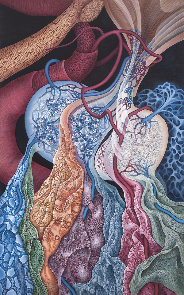
Acrylic on canvas, 32″ x 20″ Private collection, Pittsburgh, PA ©
Penny Oliver
"The Fabric of Life" is about the pituitary gland (hypophysis). This painting presents the grandeur of this tiny little gland by visually connecting so many of the organs it serves and displaying the intricate portal system. In this piece I have represented the hormones excreted by the pituitary as flowing drapes of fabric (thus the title). Each of the drapes depicts the histology of eight of the organs that rely on the pituitary for proper function. They are shown, from left to right: thyroid, adrenal gland, breast, ovary, testis, bone, kidney, uterine smooth muscle. The carotid artery sits in the background as a reminder of the importance of the blood flow to carry the hormones throughout the body. I have taken the wall of the artery off in layers to expose the smooth muscle, the elastic membrane and the endothelium. I have shown the cells of the adenohypophysis as well as the neurohypophysis (with herring bodies). The blood supply is intricately woven throughout. The hypothalamus is present in that I have shown pathways for the neurosecretory cells to travel to the posterior lobe as well as to the hypophyseal portal system.
Penny Oliver
"The Fabric of Life" parla della ghiandola pituitaria (ipofisi). Il dipinto rappresenta la grandezza di questa piccola ghiandola collegando visivamente tanti degli organi che serve e mostrando l'intricato "sistema portale". In questo pezzo ho rappresentato gli ormoni secreti dall'ipofisi come fluenti drappi di tessuto (da qui il titolo). Ciascuno dei teli raffigura l'istologia di otto degli organi che si affidano all'ipofisi per il corretto funzionamento. Sono mostrati, da sinistra a destra: tiroide, ghiandola surrenale, seno, ovaio, testicolo, osso, rene, muscolatura liscia uterina. L'arteria carotide si trova sullo sfondo come promemoria dell'importanza del flusso sanguigno per trasportare gli ormoni in tutto il corpo. Ho rimosso la parete dell'arteria a strati per esporre la muscolatura liscia, la membrana elastica e l'endotelio. Ho mostrato le cellule dell'adenoipofisi e la neuroipofisi (con corpi di aringhe). L'afflusso di sangue è intrecciato in modo intricato. L'ipotalamo è presente in quanto ho mostrato i percorsi che le cellule neurosecretorie usano per viaggiare verso il lobo posteriore e verso il "sistema portale" ipofisario.
Penny Oliver

Adenoma dell’ipofisi
Adenoma dell’ipofisi e endoscopia endonasale L’adenoma ipofisario è un tumore benigno, che colpisce l’ipofisi e in particolare la sua parte

Endoscopia ipofisaria transnasale
Adenoma ipofisario: l’intervento Per affrontare questo tumore benigno dell’ipofisi è spesso necessario prevedere un intervento chirurgico, a meno che non


